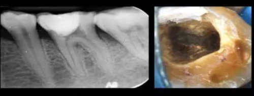
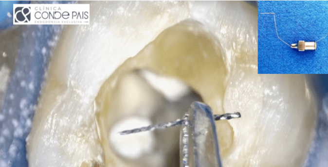
Fracture or separation of endodontic instruments is one of the main complications in our clinical practice in root canal treatment. This eventuality can negatively affect the outcome of treatment, as the blockage caused by the instrument fragments will prevent correct debridement and removal of pulp tissue, bacteria or smear layer. The prognosis in these cases will depend directly on the preoperative situation, being better in vital cases or in cases where prior disinfection has been performed.
When nickel-titanium rotary instruments (NITI) are used, the incidence of fracture ranges from 0.4% to 5%. The incidence of removal of these instruments varies widely, from 48% to 95% and is directly related to the techniques employed and the condition of the instrument. The use of ultrasonic tips and loop techniques under the surgical microscope is considered the optimal strategy for successful removal of fractured instruments. The use of the surgical microscope provides adequate visualisation and accessibility to the fractured instrument and is a key role in instrument removal.
This article describes the treatment of a 4.6 tooth with a fractured instrument and calcified metamorphosis.
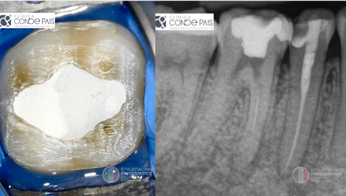
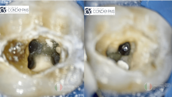
图2. 局部麻醉,隔离患牙,建立髓腔入路。使用DG16牙髓探针定位4根管,近中两根,远中两根。在近颊根管内,定位分离器械。在这个阶段,即开始使用显微镜,并利用超声修整开髓洞形。
After anaesthesia and isolation of the operative field, cavity access was performed. Four canals were located, using a DG16 endodontic explorer, two in the mesial root and two in the distal root. In the mesiovestibular canal we located the fractured instrument. In this phase we used an operating microscope (CJ OPTIK, Ablar, Germany) and redefined the access cavity with ultrasonic tips.
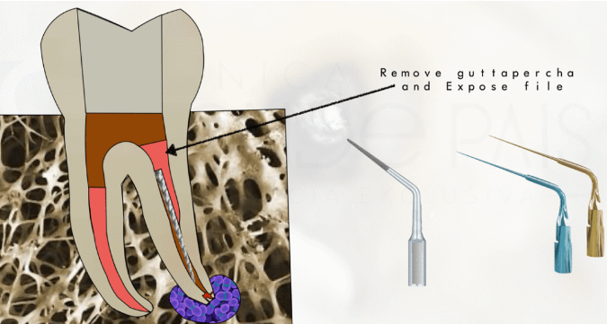
图3. 使用超声器械暴露分离器械。
We proceed to the removal of the broken file using the loop technique following this process: We expose the instrument with ultrasonic tips.
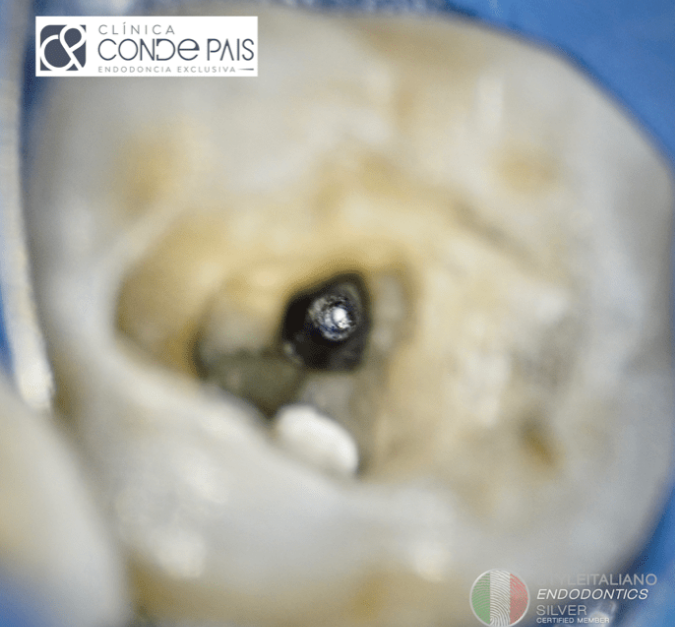
图4. 暴露的器械。
File eposed
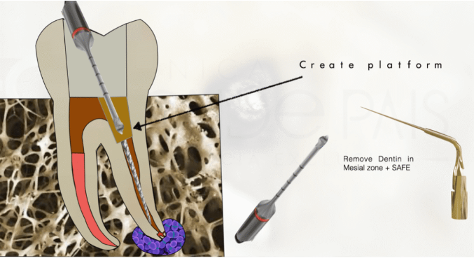
图5. 使用小直径超声工作尖在分离器械上方建立工作平台,期间使用17% EDTA溶液湿润根管。
We create a working platform over the fractured instrument with small calibre ultrasonic tips and wet canal with EDTA 17% liquid.
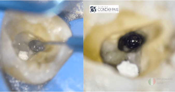
图6. 建立的工作平台。
Working Platform
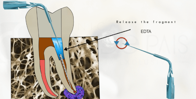
图7. 松解分离器械,并在器械周围创建至少1mm的空间。
We release the fractured instrument and created a space of at least 1 mm around the instrument.
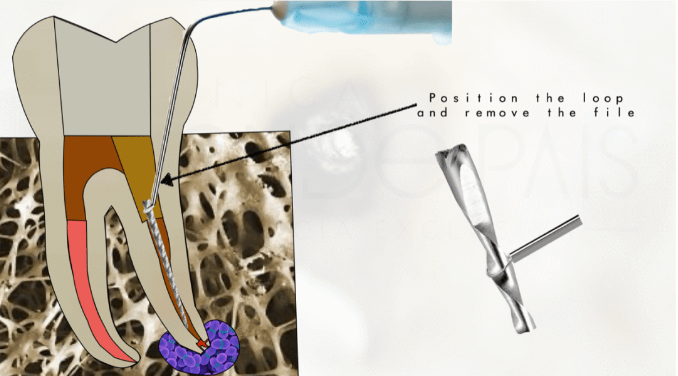
图8. 将套索绕住器械断端。
We position the loop around the instrument.

图9. 收紧套索,向上牵拉分离器械直至取出。
Close the loop and make traction movements until the broken file is removed.
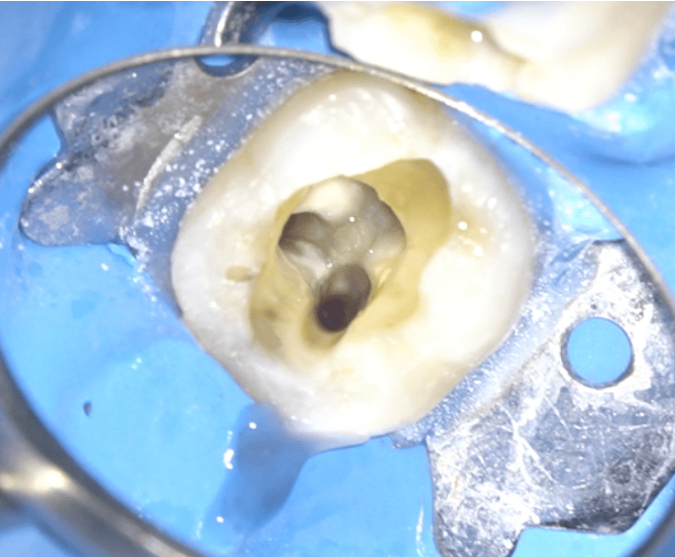
图10. 分离器械取出后,重行根管治疗。使用10#C+锉探查根管,使用根尖定位仪测量工作长度,并通过根尖片进行核对。当根管疏通顺滑后,器械预备根管至25#06锥度。更换锉间使用5.25%次氯酸钠冲洗根管。
Once the instrument fragment has been removed, we proceed with the endodontic treatment.
We performed an exploration of the root canals with C+ ISO 10 files and measured the working length using an electronic apex locator and checked it against parallel periapical radiographs.
Once a correct glide path was created, we proceeded to instrument the canals to (25-06). Between file and file we used 5.25% sodium hypochlorite as irrigant, using the IrriFlex tip for its delivery.
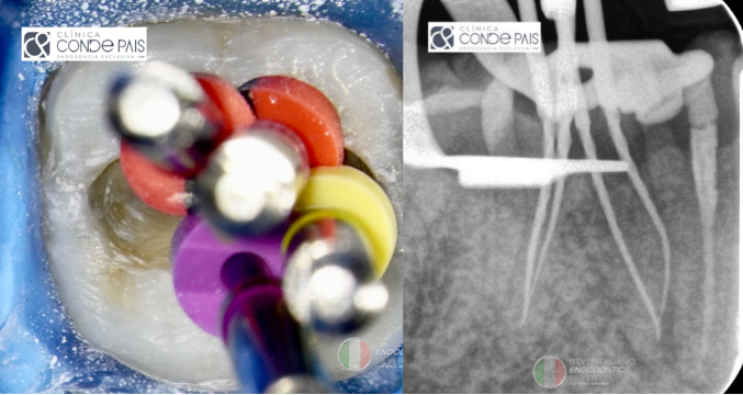
图11. 在封闭根管前核对工作长度。
We check the working length before sealing the canals.
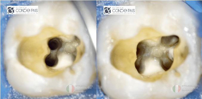
图12. 当所有的根管都器械预备完成后,确认根尖的通畅度和工作长度,对根管系统进行终末冲洗。使用5.25%次氯酸钠、生理盐水和17% EDTA。
Once all the canals have been instrumented, apical permeability and working length have been checked, we proceed to the final irrigation of the canal system. For this step we use 5.25% sodium hypochlorite, physiological saline solution and EDTA 17% solution.
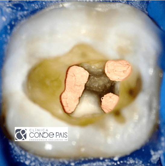
图13. 根管系统成形并消毒后,干燥根管,使用连续波热牙胶技术充填根管,确保根管系统的恰当三维封闭。
After shaping and disinfection of canal system, we proceed to dry and fill using continuous wave technique, ensuring a correct 3D seal.
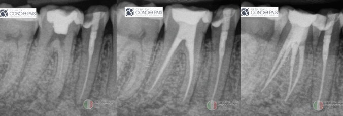
图14. 当根管充填完毕并确认无误后,继续行冠方暂时封闭,使用粘结技术和大块树脂,光固化后暂封,后行最终修复。
Once the obturation has been made and checked to be correct, we continue with the immediate sealing with adhesive technique and Bulk Fill composite, after light curing we place provisional cement and we return to the referrer for definitive restoration.
免责声明:以上文章来源于江苏牙体牙髓 ,作者闫明 译,仅供医生学习交流,如有侵权,请联系删除!


