
以下文章来源于江苏牙体牙髓 ,作者闫明 译
原文网址:https://www.styleitaliano.org/deep-resin-infiltration-for-the-conservative-management-of-a-central-incisor-affected-by-mih/
2001年Weerheijm等学者首次将磨牙-切牙釉质矿化不全(Molar-incisor hypomineralisation, MIH)定义为“全身因素引起的釉质矿化不全,表现为1~4颗第一恒磨牙局部实质性釉质缺陷,常伴切牙受累”。2003年,MIH被描述为一种由矿化和无机釉质成分降低引起的发展性实质性釉质缺陷,导致受累牙釉质变色破损。虽然病因尚不清楚,但其局限性、非对称性的病损似乎与全身因素有关。MIH的临床表现取决于其严重程度,从白色乳白色不透明斑块,黄棕色不透明斑块,萌出后釉质破损,到非典型的龋损定位于至少一颗第一恒磨牙,累及或不累及切牙。
Molar-incisor hypomineralisation (MIH) was first defined in 2001 by Weerheijm et al. and as “hypomineralisation of systemic origin, presenting as demarcated, qualitative defects of enamel of one to four first permanent molars frequently associated with affected incisors”. In 2003, MIH was described as a developmental, qualitative enamel defect caused by reduced mineralization and inorganic enamel components which leads to enamel discoloration and fractures of the affected teeth. Although the etiology remains unclear, the localized and asymmetrical lesions seem to have a systemic origin. The clinical presentation of MIH depends on its severity and can range from whitecreamy opacities, yellow-brown opacities, post-eruptive enamel breakdown, to atypical caries located on at least one first permanent molar with or without incisor involvement.
◆MIH患者常见的临床问题:
①萌出后的釉质破损导致牙本质暴露,增加牙髓受累的风险;
②牙齿敏感;
③美观问题;
④难以实施局部麻醉;
⑤牙齿缺失。
◆Common clinical problems for patients with MIH are:
①Post-eruptive enamel breakdown leading to dentine exposure which increases the risk of pulp involvement
②Tooth Sensitivity
③Aesthetic Problems
④Difficulty in administering local anaesthesia
⑤Tooth loss
虽然树脂渗透对于浅表白色斑块的轻度MIH病例可以显现良好的效果,但对于较深的病损的切牙需要改良技术,被称为“深层树脂渗透技术”。该技术包括对受累牙的粗研磨预备,无论是口内喷砂还是低速圆球钻都要确保渗透材料可以到达病损区域的全部范围。
Although resin infiltration has shown good results in mild MIH cases with superficial white lesions, deeper lesions require a modified technique for the aesthetic management of incisors, called “deep resin infiltration technique” (Attal et al.). This technique involves preparation of the affected tooth by macroabrasion, either by intraoral sandblasting or a round bur at low RPM to ensure that the infiltration can indeed reach the full extent of the lesion in case of MIH.
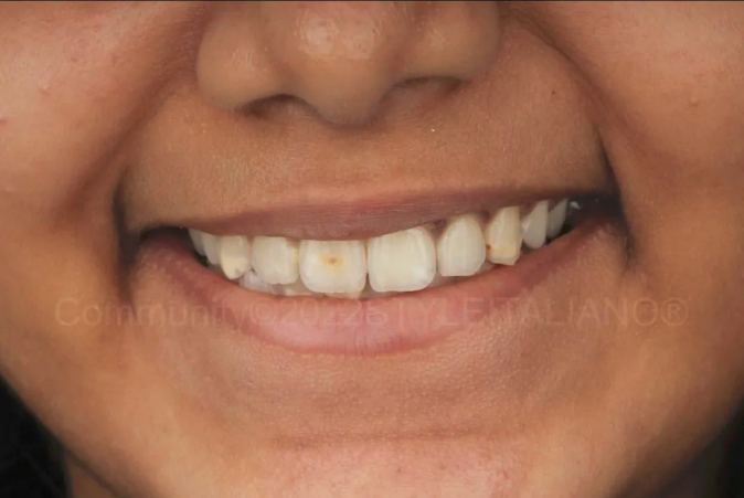
图1. 患者女,12岁,因前牙深褐色斑块就诊。她表达了在学校和社交场合因为牙齿上的棕色点被人嘲笑的尴尬,使他非常绝望。
A young girl, aged 12 came into our dental office with the chief complaint of the dark brown area on her front tooth. She expressed her embarrassment at school and social situations where she was being teased about the brown spot causing her a lot of despair.
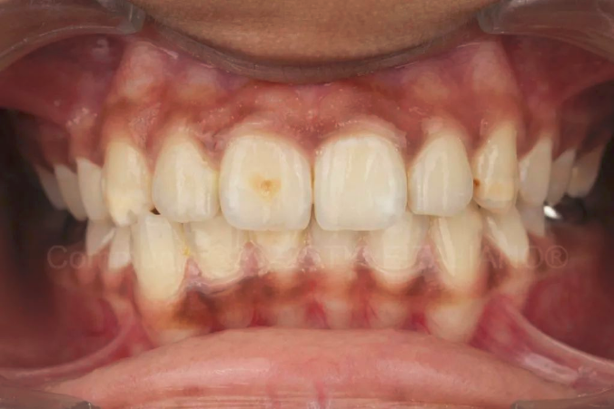
图2. 病史和检查显示11、13、23、41均有棕色变色。两颗下颌第一恒磨牙均在9岁时行根管治疗及金属冠修复。上颌恒磨牙可见矿化不良的表面上存在大面积的复合树脂修复体。根据MIH的分类,该病例属于重度,即萌出后釉质破损、牙冠损坏、累及釉质伴龋损,牙本质敏感史及美观困扰。
History and examination revealed the presence of brown discoloration of 11, 13, 23 and 41. Both lower first permanent molars were root canal treated at the age of 9 and given metal crowns. The upper permanent molars showed large composite restorations with hypomineralized tooth surfaces. According to the classification of MIH by Mathu – Muju and Wright, this case could be classified as Severe MIH, i.e. post-eruptive enamel breakdown, crown destruction, caries associated with affected enamel, history of dental sensitivity and aesthetic concerns.
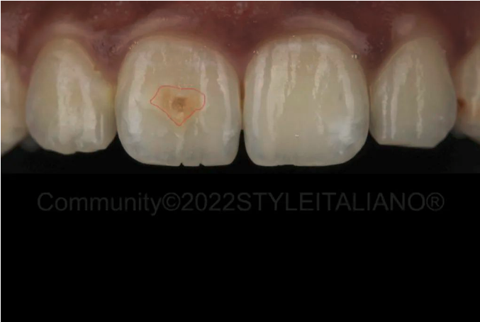
图3. 术前可见11唇面边界清晰棕色病损。因其他患牙不是患者的主诉问题,所以仅治疗11和23不连续的近中表面,该处容易发生龋病。13和41上的病损有坚硬的釉质表面,建议以后定期随访。11将进行粗研磨、深层树脂渗透和复合树脂修复治疗,23将行复合树脂修复。
Pre operative upper anterior close up reveals a well demarcated brown lesion with 11. The lesions on the other teeth were not a primary concern for the patient hence it was decided to only treat the lesion on 11 and also 23 which showed a discontinuity of the mesial surface, making it prone to caries. The lesions on 13 and 41 had a hard enamel surface and would be checked at regular follow up visits for further treatment. Tooth 11 would be treated with macroabrasion, Deep Resin Infiltration (with ICON, DMG) and composite restoration, while tooth 23 would be treated with a proximal composite restoration.
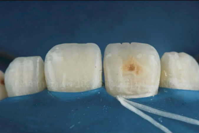
图4. 橡皮障隔离是确保出色的软组织保护和无污染的渗透和粘接环境所必须的。
Isolation with rubber dam is mandatory to ensure excellent protection of the soft tissues and too create contaminant-free environment for infiltration of the lesion and for bonding of the composite.
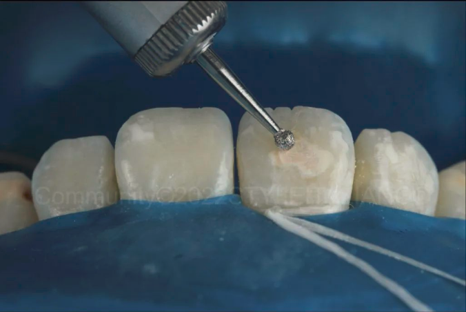
图5. 粗研磨使用慢速大球钻去除浅表釉质,使得酸可以更深层的渗透和随后的渗透可有效的掩盖变色。釉质的去除要平整、浅表,控制车针以免磨出凹坑。
Macroabrasion is done with a large round bur at slow speed to remove the superficial enamel. This allows deeper penetration of the acid and subsequently the infiltrant to effectively mask the discoloration. The enamel is removed with a very smooth, shallow and controlled movement of the bur without causing any deep pits.
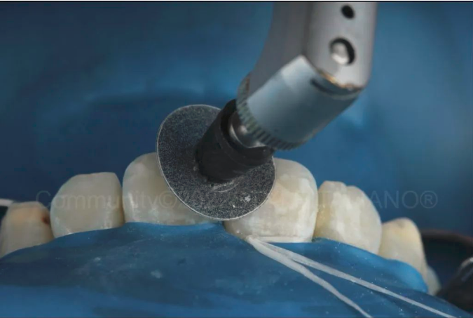
图6. 粗颗粒抛光碟平滑预备区域的边缘,可使复合树脂材料更好的过渡。
A coarse disc (Shofu Snap on Disc) is used to smoothen the margins of the preparation to allow better blending of the composite material.
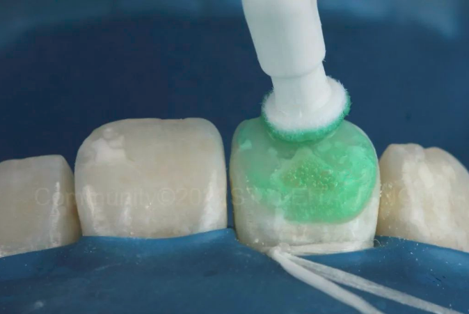
图7. 酸蚀累及区域2分钟,盐酸有助于渗透非多孔的釉质表面。
Icon Etch is applied on the affected area for 2 mins. The hydrochloric acid helps permeabilize the non porous enamel surface.
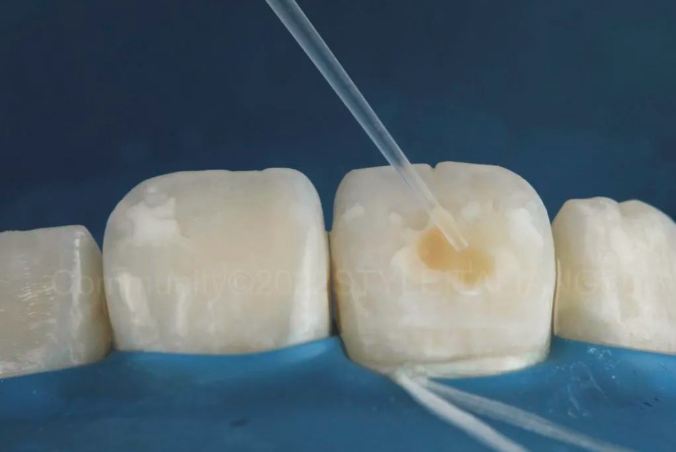
图8. 经过彻底地冲洗酸蚀剂并干燥表面后,下个步骤是乙醇干燥2分钟。该步骤可观察到树脂渗透后的效果,如果效果不满意,可以重复酸蚀最多3次。
After thoroughly washing the etching gel and drying the surface, the next step in the infiltration technique is the Icon Dry (alcohol solution). It is applied for 2 minutes in order to visualize the result achievable with resin infiltration. If the desired result is not achieved, these 2 steps can be repeated up to maximum 3 times, according to manufacturer’s instructions.
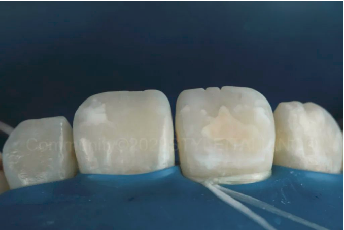
图9. 我们酸蚀干燥了两轮后决定停止。
We decided to stop after 2 cycles of Etch and Dry.
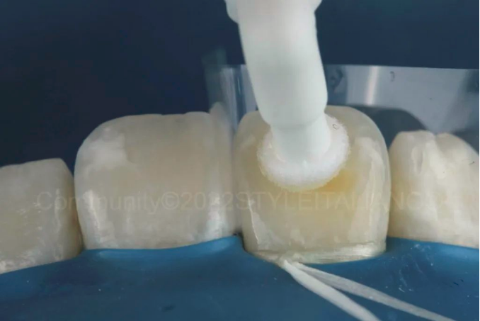
图10. 聚酯薄膜隔离患牙,树脂渗透3分钟,然后光固化40秒。然后重复渗透1分钟以上再固化。
After using a mylar strip to separate the teeth, the Icon Infiltrant is applied on the tooth surface for 3 minutess and then photocured for 40 seconds. This is repeated for 1 minute more and photocured.
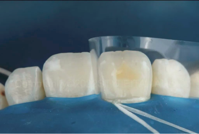
图11. 树脂渗透后的情况,仍可见少许黄色区域,将使用一层复合树脂覆盖。
This was the situation after resin infiltration. Some yellow area is still visible but it will be covered with a layer of composite resin.
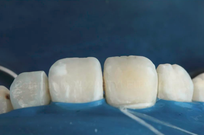
图12. 体色复合树脂遮色,修复体修整抛光。
After adding a Body shade composite (Filtek Z350Xt, 3M), the restoration was finished and polished.
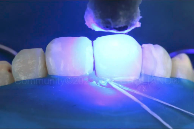
图13. 甘油层下光固化。
Photocuring under a layer of glycerin gel.
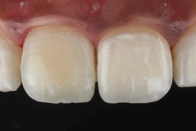
图14. 术后即刻。最终结果需要等待牙齿再水化至少48小时。
Immediate post operative result. We must always wait for rehydration of the teeth for at least 48 hours to assess the final result.
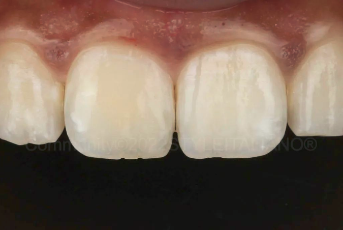
图15. 两周后的最终效果显示可接受的融合和美学。
Final situation after 2 weeks showing acceptable integration and aesthetics.
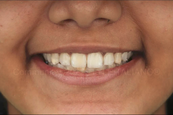
图16. 最终微笑照。
Final smile.
免责声明:本文来自于网络,仅供医生学习交流,如有侵权,请联系删除!


