以下文章来源于江苏牙体牙髓 ,作者闫明 译
原文网址:http://endodontics.styleitaliano.org/danger-zone-in-c-shaped-root-canals/
The danger zone is an area of the root canal where the primary thickness of dentin before preparation is under 1 mm. In general, the danger zone is located 4 to 6 mm below the canal chamber orifice. A danger zone can appear in the buccal root of maxillary premolars and molars and in the distal area of the mesial root and mesial region of the distal root of the mandibular molars. The cause of forming C-shaped canals and the prevalence of the danger zone in C-shaped roots is a failure of Hertwig's epithelial root sheath to fuse on the lingual or buccal root surface. If Hertwig's epithelial root sheath fails to fuse on the lingual surface, then the groove and danger zone occur on the lingual side of the root. If the root sheath fails to unite on the buccal surface-danger zone appears on the buccal side. There is almost always a danger zone in roots containing C-shaped root canals. Very little dentin separates the external surface from C-shaped canal system, thereby increasing the possibility of perforation during endodontic treatment and prosthetic restoration. (Melton et.al. JOE 1991). In 80% of C-shaped root canals in mandibular molars, invagination and correlated danger zone occurs in 80% lingually, 19% buccally, and only 1% have no groove or danger zone (Avi Shemesh, Avi Levin, Vered Katzenell, Joe Ben Itzhak, Oleg Levinson, Zini Avraham, Michael Solomonov. Clinical Oral Investigations 2017). Lingual walls in C-shaped roots are usually thinner than buccal walls at the coronal, middle, and apical third of the roots, especially at the mesial locations, compared to the danger zone in standard lower molars, which is situated 4-6 mm below the canal orifices. The lowest value of dentine thickness in C-shaped root canals was 0,26 mm (Wen Lin Chai and Yo Len Thong JOE 2004). The danger zone in the mandibular premolars with C-shaped roots was situated lingually, mesially, or mesiolingually (.Avi Shemesh, DMD, Ella Lalum, DMD, Joe Ben Itzhak, DMD, Dan Henry Levy, DMD, MSc, Alex Lvovsky, DMD,Oleg Levinson, DMD, and Michael Solomonov, DMD JOE 2020). That's why we need to plan treatment and instrumentation according to the location of DZ. In the case of the position of invagination at the lingual side, we need to avoid lingually sweeping movements. If the invagination is situated buccally, we avoid sweeping motions on the buccal side. Due to the high risk of strip perforation, it is recommended to work with instruments of small taper 02, 04, 00, for example, XP-finisher or regressed taper such DC-taper 2 H SS-white, TrueNatomy Dentsply Sirona. Knowledge of anatomy is necessary to perform successful root canal treatment. Lack of awareness about the presence, location, and value of danger zone due to incomplete diagnostic can lead to over-instrumentation and failure, such as strip perforation and vertical root fracture. Incredibly challenging are re-treatment and broken file removal cases in C-shaped root canals because of the previous preparation of the danger zone. It finds no literature about the re-treatment or broken file removal in C-shaped root canals, but cases exist. In separated file cases, bypass is preferable. In prosthetic reconstruction, custom posts are not recommended due to the high risk of perforation during preparation.
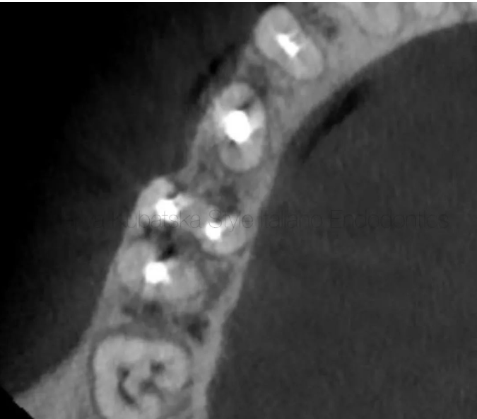
图1. 一位50岁的患者在进行根管治疗时怀疑存在两根分离器械而被转诊至我处。临床检查:冠方暂时封闭,未见明显折裂,探诊深度小于3mm,无叩痛和扪痛。牙髓状态:坏死。诊断:无症状根尖周炎。术前CBCT检查:轴向位显示C形根管C2型(C形根管的分型详见公众号早先文章《下颌第二磨牙C形根管结构》)。舌侧沟存在危险区。牙本质厚度最小值测量小于0.5mm。
50-year-old patient was referred to me with information about suspicion of separation of 2 instruments while performing a root canal treatment. On clinical examination, temporary filling IRM was detected. No signs of crack or fracture. The probing depth was no more than 3 mm. No pain on percussion or palpation. Pulp status: Necrosis. Diagnosis: Asymptomatic apical periodontitis. Pre-opp CBCT examination, the axial slice showed a C-shaped root canal type C2, according to Melton and Fan. It was noticed the presence of a danger zone in a lingual groove. The lowest value of dentin thickness according to measurement was under 0,5 mm.
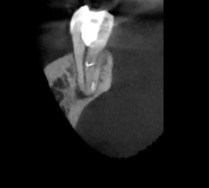
图2. 术前CBCT检查:矢状位显示在近舌根管根尖区和近颊根管冠方分别存在一根分离器械。存在根尖周病变。由于危险区定位于舌侧及其较少的牙本质厚度,决定对于近舌根管内的分离器械采取建立旁路的方法。近颊根管冠方的分离器械使用超声技术成功取出。
Pre-opp examination CBCT, sagittal slice showed the presence of a separated instrument in apical part of ML root canal and a separated instrument in the coronal part of MB root canal. A periapical lesion was present. Because of the lingual location of the danger zone and a low value of dentin sickness, I made a decision to perform a bypass in ML root canal. An instrument from the coronal part of MB root canal was successfully removed by using the ultrasonic removal technique.
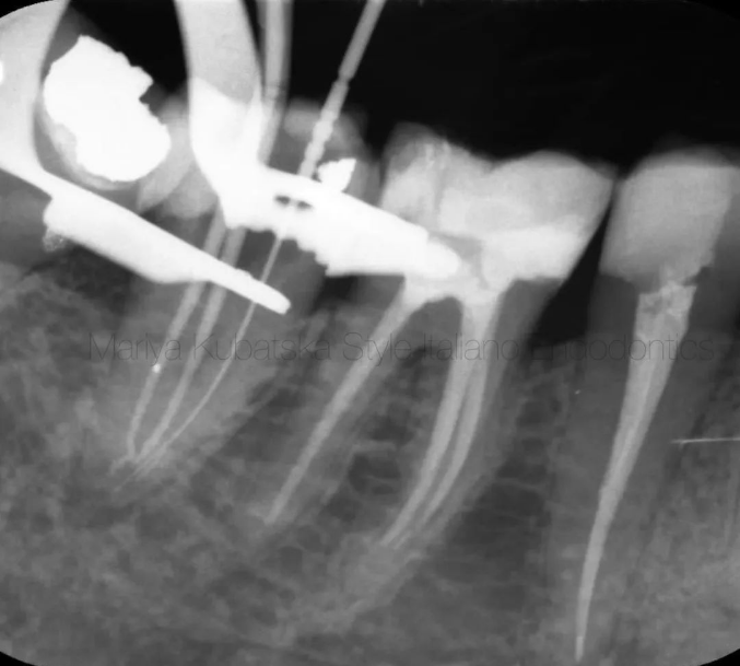
图3. 术中X线片显示:近舌根管建立了旁路,近颊和远中根管达到了工作长度。旁路使用手用C锉建立,从08#预备至20#,然后使用机用器械。三根管均预备至30#06锥度。根管冲洗使用的VDW EDDY声波荡洗和XP finisher根管锉。
Intra-op X-ray showed performed bypass in ML root canal and achieved working length in MB and D root canals. Bypass was performed by using hand files (C-pilot) from ISO 08 to 20 and machine files DC-taper 2H SS-white. ML, MB, and D root canals were instrumented to ISO 30.06 by DC-taper 2H SS-white. Endodontic solutions were activated by EDDY VDW and XP-endo finisher FKG Dentaire.
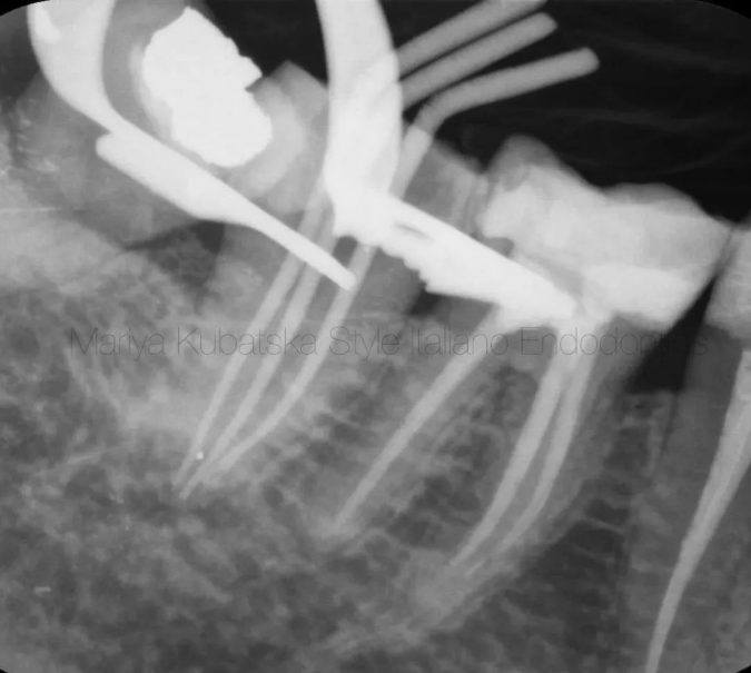
图4. 术中试尖片显示所有根管均达到合适的工作长度。
Intra-op X-ray with gutta-percha points showed correct working length in all root canals.
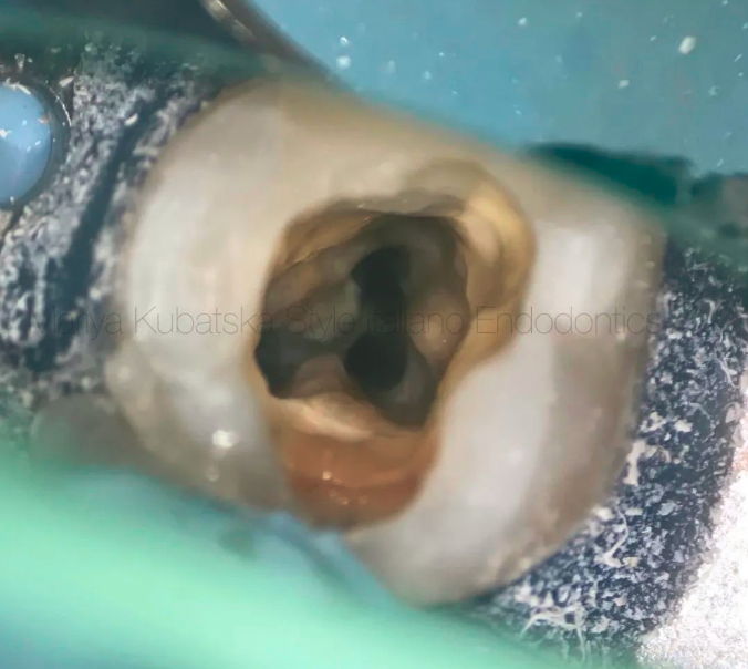
图5. 根管成形和清理后的髓室情况。
The appearance of the pulp chamber after the phases of shaping and cleaning.
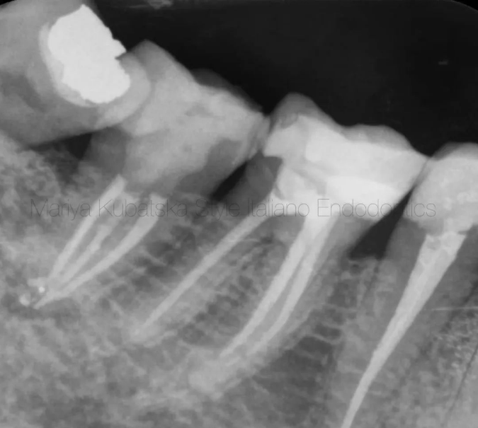
图6. 术后X线片。根管充填使用的环氧树脂糊剂和牙胶。充填方法结合侧方加压和垂直加压,根尖区使用侧方加压,剩余部分使用垂直加压。
Post operative x-ray. Obturation was made with epoxy sealer and gutta-percha by using hybrid technique. The hybrid technique is a combination of lateral and vertical compaction. The apical part of the canal is filled using the lateral compaction method, the remaining part using the vertical compaction method.
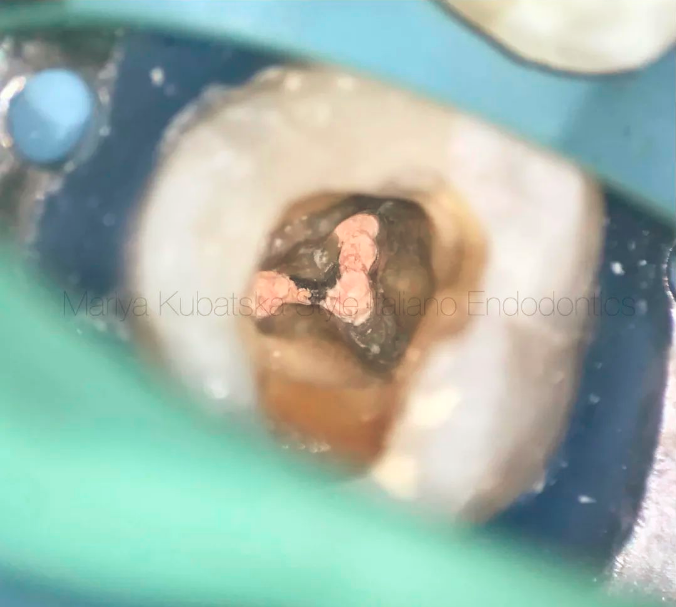
图7. 根管充填后的髓室观。
The appearance of the pulp chamber after root canals obturation.
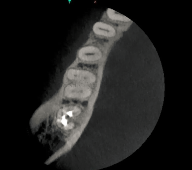
图8. 一位40岁的患者因47行非手术再治疗转诊至我处。临床检查:MOD树脂充填物,无明显折裂,探诊深度小于3mm,无叩痛及扪痛。诊断及牙髓状态:根管治疗后,无症状根尖周炎。术前CBCT检查:轴向位显示C形根管C2型。舌侧沟存在危险区。牙本质厚度测量最少小于0.3mm。怀疑先前的治疗发生带状穿孔。
A 40-year-old patient was referred to me for non surgical re-treatment of tooth 47. On clinical examination MOD composite filling was detected. No signs of crack or fracture. The probing depth was no more than 3 mm. No pain on percussion or palpation. Diagnosis and pulp status: Previously treated , asymptomatic apical periodontitis. Pre-opp CBCT examination, the axial slice showed a C-shaped root canal type C2, according to Melton and Fan. It was noticed the presence of a danger zone in a lingual groove. The lowest value of dentin thickness according to measurement was under 0,3 mm. It was suspicion of strip perforation during previous treatment.
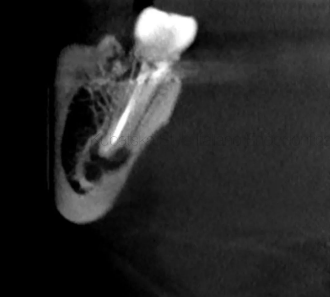
图9. 术前CBCT检查:矢状面显示存在根尖病变。47的牙根距离下颌神经管很近,这有可能会导致根管治疗的并发症。
Pre-opp examination CBCT, sagittal slice showed the presence of periapical lesion. The root of tooth 47 was found to be very close to the inferior alveolar nerve, what could cause complications during root canal treatment.
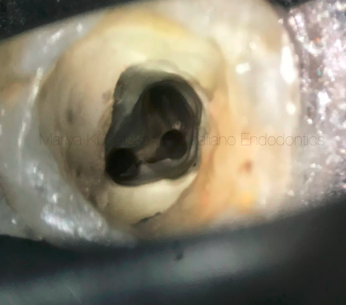
图10. 根管成形和清理后的髓腔观。牙胶使用再治疗旋转器械、XP shaper和橘油通过机械的方法去除。ML、MB和D根管均预备至30#06锥度。由于距离下颌神经管很近,出现并发症的风险较高,预备时特别注意保护根尖止点。没有探查到带状穿孔。根管冲洗使用的VDW EDDY和XP finisher。
The appearance of the pulp chamber after the phases of shaping and cleaning. Gutta-percha was removed mechanically using instruments Endostar Re Endo Rotary System, XP-endo shaper FKG Dentaire and Orange Guttane Cerkamed. ML, MB, and D root canals were instrumented to ISO 30.06 by DC-taper 2H SS-white. Due to its close location to the inferior alveolar nerve and high risk of complications was performed apical stop. Strip perforation wasn't detected, which was suspected earlier. Endodontic solutions were activated by EDDY VDW, XP-endo finisher FKG Dentaire.
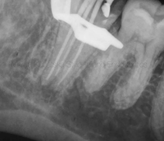
图11. 术中试尖片显示所有根管均到达合适的工作长度。
Intra-op X-ray with gutta-percha points showed correct working length in all root canals.
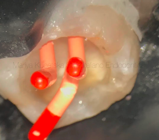
图12. 校准主尖。
Photo of calibrated gutta-percha points in root canals. Calibration was made by calibration ruler Dentsply Sirona.
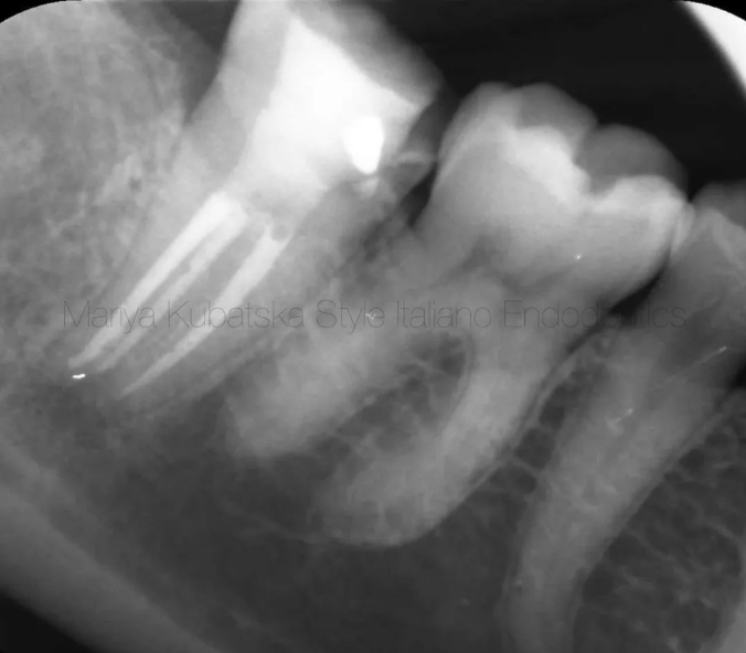
图13. 术后X线片。根管充填使用的环氧树脂糊剂和牙胶。
Post operative x-ray. Obturation was made with epoxy sealer and gutta-percha by using hybrid technique.
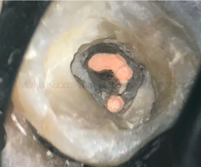
图14. 根管充填后的近中和远中根管口。
A view of the mesial and distal orifices after obturation.
总结
C-shaped root canals can be very demanding for clinicians or operators due to complex anatomy and the appearance of danger zones. RCT of C-shaped root canals containing invaginations (grooves) can be challenging in every stage of treatment, from diagnosing, instrumentation, and obturation to post-endodontic restoration. That's why deep knowledge and awareness of anatomy are mandatory. Minimal invasive instrumentation is crucial to preserve dentin, which impacts tooth functionality and long-term prognosis.
以上文章来源于江苏牙体牙髓 ,作者闫明 译
原文网址:
http://endodontics.styleitaliano.org/danger-zone-in-c-shaped-root-canals/免责声明:本文来源于网络,仅供医生学习交流,如有侵权,请联系删除!


