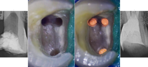
Proper clinical practice in Endodontics requires careful and detailed knowledge on the know how of using different instruments, tools and devices used in the treatment course. Even in the simplest cases, violating the limitation of some instruments or misusing/misplacing those instruments may lead to severe iatrogenic errors which may act as a barrier against providing the proper standard of endodontic care.
The aim of this article is to present a case of previously treated maxillary central incisor with a radiopaque foreign body and symptomatic apical periodontitis.
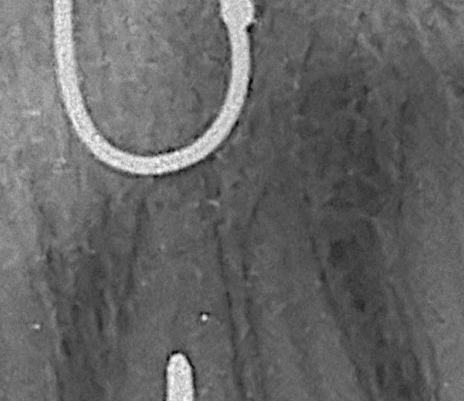
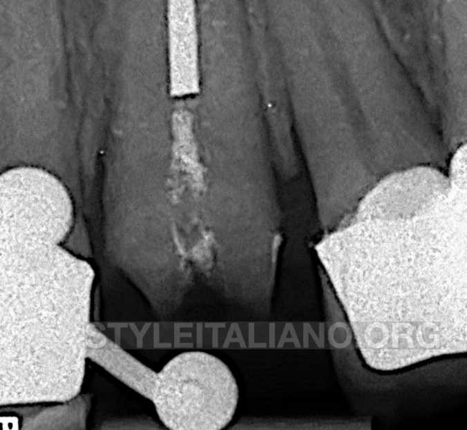
图1. 术前评估。
Pre-operative evaluation
29 years old male presented complaining of dislodged PFM crown from a maxillary central incisor which has been sensitive to biting for couple of weeks before crown loss. Clinical examination revealed a previously treated and prepared maxillary central incisor that is tender to mild percussion. Radiographic examination revealed a radiopaque fragment in the middle portion of the root canal. Apical to the fragment, the canal didn’t seem to be prepared.
A diagnosis of a pulpless, previously treated tooth with apical periodontitis was finally recorded
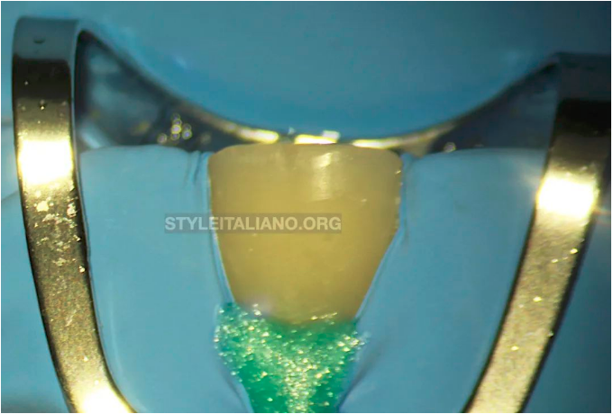
图2. 根管治疗前的冠方重建与隔离。
Pre-endodontic build up and isolation
Due to the crown mutilation and the possibility of mutli-visit treatment, a decision was taken to execute a proper pre-endodontic build up to maintain acceptable esthetics during the treatment period and to aid in the otherwise complicated isolation of the tooth
Primary isolation was done using 212 SA clamp and heavy rubber sheet. Secondary isolation was done using a light curable gingival barrier.
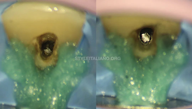
图3. 建立至折断片的直线通路。
Straight line access to the fragment
Under high microscopic magnification, straight line visual access to the fragment was achieved by removing all overhanging dentin palatally. This was done by refining the previously attempted access using diamond coated ultrasonic tip.
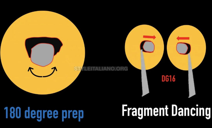
图4. 计划取出异物折断片。
Planning retrieval of the foreign fragment
Due to the cross-section of the coronal part of the root canal in a maxillary central Incisor, it was assumed that fragment is engaging dentin palatally [in the narrow part] while being free in the labial direction [wide part]. This was confirmed by inserting a hand #20 k-file bypassing labial to the fragment.
The plan was to prepare a 180 degrees preparation to a depth of 1-2 mm lingual to the fragment until visual confirmation of looseness is evident
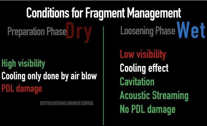
图5. 折断片取出的条件。
Conditions for fragment management
It has to be highlighted that different stages of management dictate the use of different conditions and media.
During preparation phase, a dry condition was used to provide good visibility, where cooling during preparation is done using a stropko tip blow. It is important to adopt the mother in law rule [10 second rule] during the preparation phase to avoid periodontal damage
During the loosening phase, wet condition is used to provide a cavitation effect and acoustic streaming that will aid in loosening
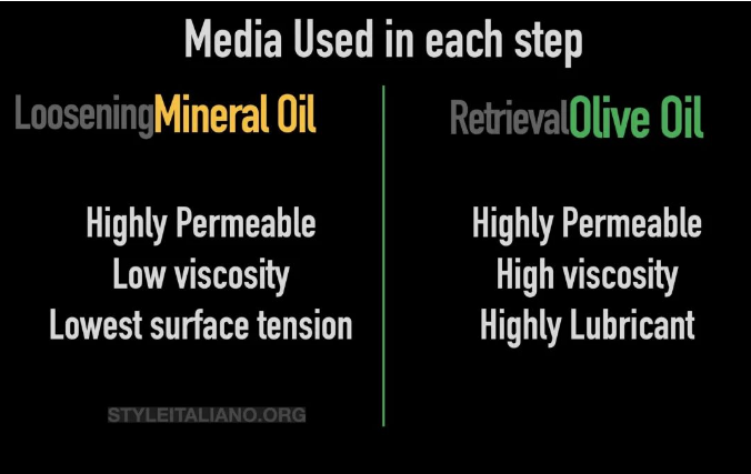
图6. 取出各阶段使用的介质。
Type of media used in each step of management:
During the loosening phase, Mineral oil [Johnson’s baby oil] was used as it permeates beneath the fragment well owing to low surface tension and viscosity
During the retrieval attempt, based on the recommendation of Dr. Yoshi Terauchi, olive oil has been used owing to its high lubrication potential as well as its high viscosity.
超声激活尝试取出折断片
Retrieval attempts using Ultrasonic activation
Once the fragment become dancing in the root canal space, Olive oil was used to fill the pulp chamber to the cavo-surface margin. An ultrasonic tip was activated in the preparation site and labially [alternatively] until the fragment popped out from the root canal. It appeared that the fragment was a broken Endo-Z bur.
图7. 观察确认。
Visual Confirmation
After retrieval, high magnification was used to confirm that the root canal space is free from any impediment.
Canal preparation was done using reciprocating files [45/.05]
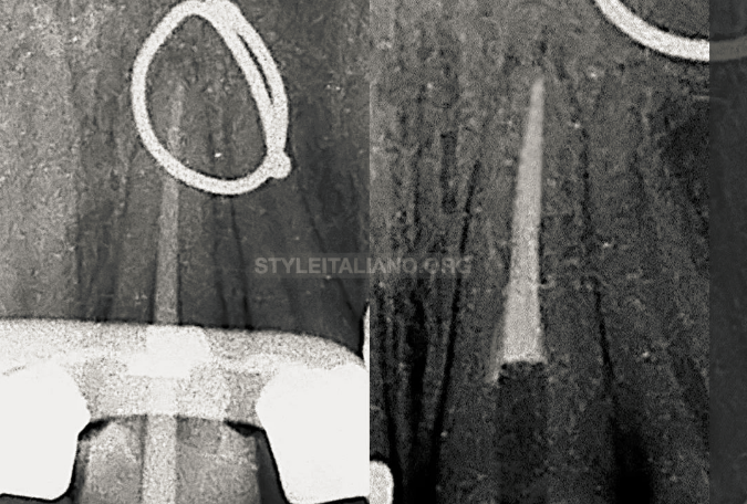
图8. 充填。
确定合适主尖后,根管使用热牙胶垂直加压充填。
Obturation:
Following cone fitting procedures, the root canal was obdurated using GP and resin sealer using warm vertical compaction technique.
免责声明:以上文章来源于江苏牙体牙髓 ,作者闫明 译,如有侵权,请联系删除!


